
Selected works
TopoMortar: A dataset to evaluate image segmentation methods focused on topology accuracy
in Arxiv, 2025.
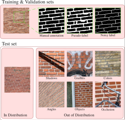
TopoMortar is the first dataset specifically designed to evaluate whether topology-enhancing methods do actually improve topology accuracy. TopoMortar allows to portray different challenging scenarios where, unlike in existing datasets, an increase in topology accuracy can be attributed to the incorporation of prior topology knowledge.
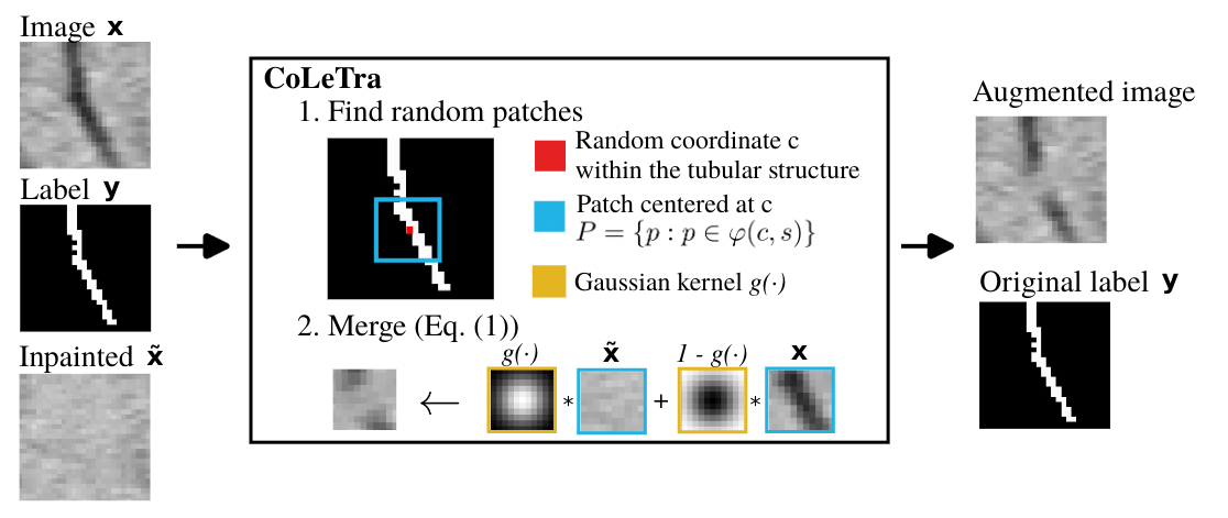
Disconnect to Connect: A Data Augmentation Method for Improving Topology Accuracy in Image Segmentation
in Arxiv, 2025.
Sauron U-Net: Simple automated redundancy elimination in medical image segmentation via filter pruning
in NeuroComputing, 2024.
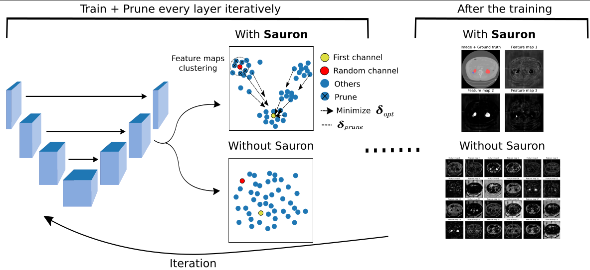
SauronUNet is a filter pruning method for UNet-like architectures that, first, increases the redundancy among feature maps to, later, eliminate them.
[ArXiv], [Code], [![]() Publication]
Publication]
Region-wise Loss for Biomedical Image Segmentation
in Pattern Recognition, 2023.
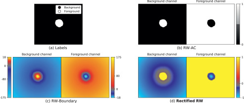
Region-wise loss is a loss function that allows you to penalize pixels based on the distances to the structures' boundary.
[ArXiv], [Code], [![]() Publication]
Publication]
Transfer Learning in Magnetic Resonance Brain Imaging: A Systematic Review
in Journal of Imaging, 2021.
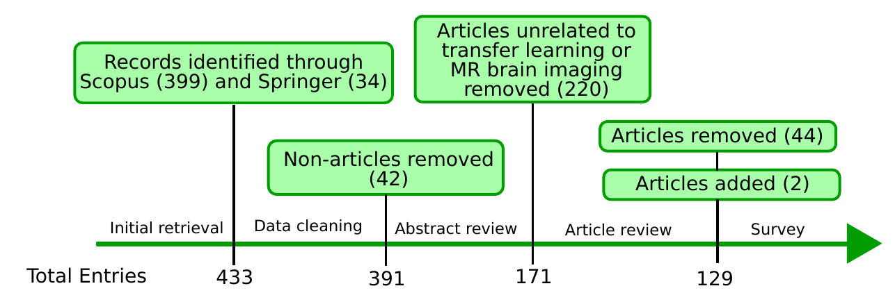
In this review, we identify research directions, gaps in knowledge, applications, and widely used strategies among the transfer learning approaches applied in MR brain imaging.
[ArXiv], [![]() Publication]
Publication]
RatLesNetv2: A Fully ConvNet for Rodent Brain Lesion Segmentation
in Frontiers in Neuroscience, 2020.
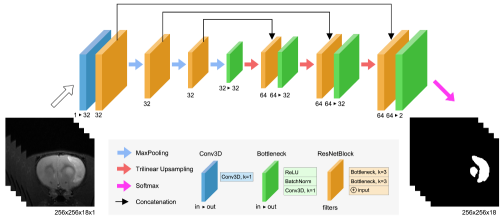
RatLesNetv2 is a convolutional neural network to segment rodent brain lesions in MR images that we validated on an exceptionally large dataset of 916 T2-weighted rat brain MRI scans.
[ArXiv], [Code], [![]() Publication], [Cake]
Publication], [Cake]
Full publications list
 Open access
  Preprint available
  Code
Code
Submitted preprints
- J.M. Valverde, M. Koga, N. Otsuka, A.B. Dahl. TopoMortar: A dataset to evaluate image segmentation methods focused on topology accuracy
- J.M. Valverde, M. Østergaard, A. Rodriguez-Palomo, P.A.S. Vibe, N.K. Wittig, H. Birkedal, A.B. Dahl. Disconnect to Connect: A Data Augmentation Method for Improving Topology Accuracy in Image Segmentation
- J.M. Valverde, V. Imani, J. Tohka. Fine-tuning ImageNet-pretrained models in medical image classification: Reassessing the impact of confounding factors
Journal papers
- J.M. Valverde, A. Shatillo, J. Tohka. Sauron U-Net: Simple automated redundancy elimination in medical image segmentation via filter pruning.
In NeuroComputing, 2024.
- J.M. Valverde, J. Tohka. Region-wise Loss for Biomedical Image Segmentation.
In Pattern Recognition, 2023.
- J.M. Valverde, A. Shatillo, R. De Feo, J. Tohka. Automatic cerebral hemisphere segmentation in rat MRI with ischemic lesions via attention-based convolutional neural networks.
In NeuroInformatics, 2022.
- R. De Feo, E. Hämäläinen , E. Manninen, R. Immonen, J.M. Valverde, X.E. Ndode-Ekane, O. Gröhn, A. Pitkänen, J. Tohka. Convolutional Neural Networks Enable Robust Automatic Segmentation of the Rat Hippocampus in MRI After Traumatic Brain Injury.
In Frontiers in Neurology, 2022.
- J.M. Valverde, V. Imani, A. Abdollahzadeh, R. De Feo, M. Prakash, R. Ciszek, J. Tohka. Transfer Learning in Magnetic Resonance Brain Imaging: A Systematic Review.
In Journal of Imaging, 2021.
- R. De Feo, A. Shatillo, A. Sierra, J.M. Valverde, O. Gröhn, F. Giove, J. Tohka. Automated joint skull-stripping and segmentation with Multi-Task U-Net in large mouse brain MRI databases.
In NeuroImage. 2021
- J.M. Valverde, A. Shatillo, R. De Feo, O. Gröhn, A. Sierra, J. Tohka. RatLesNetv2: A Fully ConvNet for Rodent Brain Lesion Segmentation.
In Frontiers in Neuroscience, 2020.
Refereed conference papers
- J.M. Valverde, A. Shatillo, R. De Feo, O. Gröhn, A. Sierra, J. Tohka. Automatic Rodent Brain MRI Lesion Segmentation with Fully Convolutional Networks.
In International Workshop on Machine Learning in Medical Imaging, 2019.
- J.M. Valverde*, V. Imani*, J.D. Lewis, J. Tohka. Predicting intelligence based on cortical WM/GM contrast, cortical thickness and volumetry.
In Challenge in Adolescent Brain Cognitive Development Neurocognitive Prediction, 2019.
International abstracts
- V. Imani, J.M. Valverde, M. Prakash J.D. Lewis, O. Gröhn, J. Tohka. Fluid Intelligence Classification Based On Cortical WM/GM Contrast, Cortical Thickness and Volumetry. In Organization for Human Brain Mapping, 2020.